Friday, December 30, 2016

First Wireless, App-Powered Handheld Ultrasound gets FDA approval.

clarius probes


Mobile Ultrasound developed by Clarius Mobile Health got the FDA 510(k) clearance for its C3 and L7 Clarius wireless ultrasound scanners on December 14, 2016.It has been rated as top technical innovation of 2016, that can change the medical device industry.

Founded in 2014 by Laurent Pelissier, Clarius Mobile Health merges the power of mobile phones, advanced technology, and decades of collective ultrasound experience to produce high quality, point-and-shoot mobile ultrasound devices.

Clarius is based at VANCOUVER, British Columbia.

The device uses just about any iOS or Android phone or tablet as the display. A Clarius app is used to control the transducers and displaying the visualizations.


Clarius’ultrasound scanners are mobile devices compatible with Apple iOS and Google Android devices, the company said.[i]

The scanners are powered by a rechargeable battery that  will last for more than 45 minutes of scanning and up to 7+ days of stand-by power. 

Two batteries and a charger are delivered with each Scanner. Built with a magnesium case, Clarius Scanners are designed to withstand challenging environments and are water submersible for easy cleaning and disinfection.

With automated gain and frequency settings it is as easy to use as a mobile camera.


"Physicians have been asking for a portable ultrasound system that works with their iPhone for some time," said Laurent Pelissier, Chairman and CEO of Clarius Mobile Health.  "The challenge has been to make an affordable device that is small enough to carry around and that also produces great images.  I'm happy to say we've succeeded in creating a product that will enable more clinicians to use ultrasound anywhere to improve patient care. It's as easy to use as a mobile phone camera."

2016 was the 200th anniversary of the stethoscope and every doctor will eventually hold a hand-held ultrasound that can be used in daily practice, just like a stethoscope  believes Laurent Pelissier, CEO of Clarius. 


"Clarius is the future of patient care. The image quality is amazing for any scanner, much less one that fits in my pocket," said Dr. Steven Steinhubl, Director of Digital Medicine at the Scripps Translational Science Institute.  "The ability to wirelessly connect it to any Apple or Android device means that anyone on my team can use it with whatever they already carry around in their pocket."

This mobile ultrasound startup is reshaping the $ 6 billion healthcare market. [ii]

In the United States, Clarius Ultrasound Scanners start at $6900.

Clarius will be available with Color Doppler on its premium offering, which will be priced higher than its entry-level black and white model.


[i] http://www.massdevice.com/clarius-wins-fda-510k-wireless-ultrasound-device/
[ii] http://www.forbes.com/sites/julianmitchell/2016/12/27/this-mobile-ultrasound-startup-is-reshaping-a-6-billion-healthcare-market/#4edb7bda4941

Women at a Dutch IVF clinic fertilized by wrong sperms by mistake.


26 women may have been fertilized by wrong sperm from a man other than their partner at the Utrecht University Medical Centre (UMC). The officials admitted it to be a ‘procedural mistake’ in a news release on  Wednesday, December 28,2016.

A technician error resulted in mix-up of sperms in fertility treatments that took place between mid-April 2015 and mid-November 2016.

The ‘mix-up’ happened in women receiving Intracytoplasmic Sperm Injection (ICSI) treatment. UMC says that although the possibility is small, it cannot be totally ruled out.

"During fertilization, sperm cells from one treatment couple may have ended up with the egg cells of 26 other couples," it said.

One of the lab technician used an inappropriate pipette to inject sperm. During each procedure of Sperm Injection, he changed the pipette but used the same rubber top, until he found traces of sperms inside the top and sounded the alarm. Usually the rubber top comes with a filter, but in these cases the top did not have a filter.  

Out of the 26 couples involved, 9 patients have already delivered, 4 have an ongoing pregnancy and the rest 13 embryos were frozen.

The couples have been informed about the mix-up and they will be meeting the doctors very shortly. They are offered DNA test to determine the paternity.

UMC said “it will do everything within its powers to give clarity on the issue as soon as possible."
Nearly 700 ICSI procedures are performed each year at the UMC.

Mix-up in fertility treatments are rare, a Singapore mother in 2012 sued the fertility clinic when her baby boy born after treatment at the clinic had markedly different features as compared to her Caucasian husband.


Sources:
UMC news bulletin
CNN news
The telegraph
BBC
The Australian.


Thursday, December 29, 2016

USPSTF advises against the routine serological screening for Herpes in teens and adult.


https://c1.staticflickr.com/9/8233/28295539863_f00c68a306_b.jpg

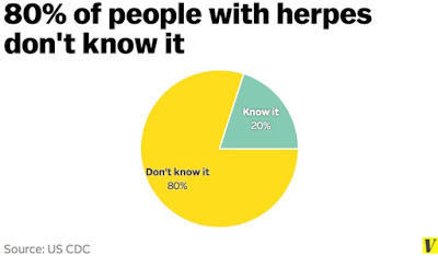
The WHO estimated that half a billion people worldwide aged 15-49 years have genital infection caused by Herpes virus. In USA, nearly one out of six people aged 14 to 49 years have it.[1]
Globally, WHO estimated that two-thirds of the population under 50 are infected with herpes simplex virus type 1.[2]


plannedparenthood.org

Genital herpes is caused by two type of viruses, Herpes simplex type1(HSV-1) and Herpes Simplex type 2 (HSV-2), a virus that almost exclusively causes genital infections.  HSV-1 is a closely related virus that causes both oro-labial herpes (“cold sores”) and genital herpes.[3]

The US Preventative Services Task Force (USPSTF) released an evidence statement and updated it’s 2005 recommendation on serological screening for genital herpes online on December 20, 2016 in JAMA and its own website.

The USPSTF recommends against routine serologic screening for genital herpes simplex virus (HSV) infection in asymptomatic adolescents and adults, including those who are pregnant.

Screening is indicated for certain high risk groups like men who have sex with men and HIV positive individuals.

After thorough review of evidence, USPSTF concluded that harms of screening outweigh the benefits because the current serological test used gives large number of false positive results and no confirmatory test is widely available.

CDC.org

Ann E. Kurth, PhD, MPH, a task force member said in a USPSTF news release “Despite genital herpes being common, testing is not generally helpful for people without symptoms, in part because early identification does not improve a person's health as there is no cure for herpes.” She further added “In addition, because current screening methods are often inaccurate, harms of screening include high false-positive rates and potential anxiety and disruption of personal relationships related to diagnosis."

Currently HerpeSelect® (Focus Diagnostics, Cypress, CA) is the most widely used FDA approved enzyme immunoassay for the qualitative detection of type specific HSV-2 IgG with sensitivity of 97 to 100% and specificity of 72 to 91%.

"Given the test characteristics of the most widely used serologic screening test for HSV-2 and a population infection prevalence of 15 percent, screening 10,000 people would result in approximately 1,485 true-positive and 1,445 false-positive results," the final recommendation statement said.[4]
False positive results lead to emotional and mental trauma along with unnecessary treatment with antiviral medications.

Diagnostic testing is indicated in persons who have recurrent HSV infection or belong to certain high risk groups like persons living with HIV infection.

The task force clarified its stand about HSV-1 infection, noting that although HSV-1 infection can be identified by serologic tests, the tests cannot determine if the site of infection is oral or genital.

The USPSTF recommends intensive behavioral counseling interventions to reduce the likelihood of acquiring a sexually transmitted infection for all sexually active adolescents and for adults at increased risk. The USPSTF has also issued recommendations on screening for other sexually transmitted infections, including chlamydia and gonorrhea, hepatitis B virus, HIV, and syphilis.

The American Academy of Family Physicians, the American College of Obstetricians and Gynecologists (ACOG), and the CDC do not recommend routine serologic screening for genital HSV infection in asymptomatic adolescents or adults.

The AAFP released its own recommendations that mirror the USPSTF statement.
In an accompanying editorial in JAMA by  Edward W. Hook III, MD, School of Medicine, Department of Microbiology, The University of Alabama at Birmingham, calls for improving performance of new and currently available tests.

He also acknowledged the lack of progress in test performance since the last USPSTF update in 2005. “The current USPSTF recommendation should serve to renew efforts to develop better tests for HSV, to improve management strategy, and to address the pervasive and harmful stigma associated with genital herpes," he concludes.


Link to the full article -http://jamanetwork.com/journals/jama/fullarticle/2593575
Link to the accompanying  editorial -http://jamanetwork.com/journals/jama/article-abstract/2593550


[1] https://www.cdc.gov/std/herpes/stdfact-herpes.htm
[2] http://www.who.int/mediacentre/news/releases/2015/herpes/en/
[3] http://jamanetwork.com/journals/jama/article-abstract/2593550
[4] http://www.aafp.org/news/health-of-the-public/20161221uspstfgenitalherpes.html

Tuesday, December 27, 2016

Maternal vaccines uptake during pregnancy remains low despite years of efforts.


courtesy istock. 

The development of new vaccines and the safety data now available for old vaccines ensures that more and more patients should receive the required vaccination in pregnancy. This is important in disease prevention and transfer of passive immunity to the unborn child.

The uptake of two commonly recommended influenza and the tetanus-diphtheria-acellular pertussis (Tdap) vaccine remains low despite being advised since years. The most recent national data was presented at 47thNational Immunization Conference (NIC) hosted by CDC in Atlanta, Georgia.

The data showed that Tdap vaccination coverage is only 10% in pregnancy while the influenza vaccination rate among pregnant women is about 50%, with 14% of women being vaccinated in the 6 months before pregnancy and 36% during pregnancy.

In a survey sent out by Sean O’Leary, MD, of the department of pediatrics, section of infectious diseases, at the University of Colorado, Denver and his colleagues, it was seen that only 75% of gynecologists routinely administered the Tdap vaccine, and 85% routinely administered the influenza vaccine to their pregnant patients.

Reimbursement-related issues topped the list of barriers, while lack of time during the antenatal visits, refusal by the patients and inability to maintain the stock of vaccines were other issues.

Dr O’Leary  said “Immunization delivery in the ob.gyn. setting may present different challenges than more traditional settings for adult vaccination, such as family medicine or internal medicine offices.”

Vaccines recommended for all pregnant women

The Advisory Committee on Immunization Practices (ACIP) of the Centers for Disease Control and Prevention (CDC) recommends a dose of Tdap during each pregnancy, irrespective of the patient’s prior history of receiving Tdap. Optimal timing for Tdap administration is between 27 weeks and 36 weeks of gestation to maximize the maternal antibody response and passive neonatal transfer.

Influenza vaccination is recommended if patient is pregnant during the flu season.

courtesy: shot of prevention 


Vaccines recommended for selected pregnant women who are at risk or  traveling to high risk areas.

Hepatitis A: The vaccine carries no known risks to the developing fetus.  
Hepatitis B: vaccine can be given in pregnancy in some circumstances. Limited data suggest that developing fetuses are not at risk for adverse events when hepatitis B vaccine is administered to pregnant women. [1]
Pneumococcus: safe if given in second and third trimester.[2]

General principles of Immunization in Pregnancy

The ACOG recommends routine assessment of immunization status of all pregnant women and administration of indicated vaccines.

Robust data and growing body of evidence demonstrate that administering inactivated virus or bacterial vaccines or toxoids is safe during pregnancy.

Live attenuated vaccines (eg, measles-mumps-rubella [MMR], varicella, and live attenuated influenza vaccine) do pose a theoretical risk (although never documented or proved) to the fetus and generally should be avoided during pregnancy.

Women who have inadvertently received immunization with live or live-attenuated vaccines during pregnancy should not be counselled to terminate the pregnancy because of a teratogenic risk.

Women who are breastfeeding can still be immunized (passive-active immunization, live or killed vaccines)


[1] https://www.cdc.gov/vaccines/pregnancy/hcp/guidelines.html
[2] http://www.uptodate.com/contents/vaccination-during-pregnancy-beyond-the-basics

Monday, December 26, 2016

PCOS is often underdiagnosed as the common cause of Abnormal Uterine Bleeding in Adolescents.

Image courtesy: University of Utah.
 Abnormal uterine bleeding(AUB) is very frequent in adolescents and generally lasts for 4-5 years after menarche. It is an important cause of visit to emergency room or healthcare provider in pediatric patients. Although DUB due to immaturity of hypothalamic pituitary ovarian (HPO) axis is a common cause of AUB in healthy adolescent, it is also important to rule out other pathological causes.

PCOS as a cause of AUB in adolescent’s patients is often underdiagnosed and poses a diagnostic dilemma as normal pubertal changes like acne, menstrual irregularities and hyperinsulinemia can mimic several features of PCOS.

Prompt diagnosis and treatment of PCOS is very important because of future reproductive and metabolic repercussions.[1] Evidence suggests that adolescents diagnosed with PCOS have elevated risk of Metabolic Syndrome (MetS) and premature cardiovascular dysfunction and cardiovascular disease.[2]

Adolescents with AUB are mostly managed as outpatients but some require hospitalization because of hemodynamic instability. A recent paper published in Journal of Pediatricand Adolescent Gynecology evaluated the most common etiology for AUB in hospital admitted adolescent patients with severe anemia.[3]

This retrospective study was conducted by Dr. Sofya Maslyanskaya, Assistant Professor of Pediatrics, Albert Einstein College of Medicine, Bronx, New York and her colleague at Children's Hospital at Montefiore in New York City.

The researchers identified 125 females aged 8 to 20 years admitted to the hospital for anemia with AUB from January 2000 to December 2014.

As per hospital protocols, all the subjects underwent hormonal testing for PCOS and other endocrinal disorders. Hence the data could be accessed and reviewed by the researchers for laboratory test results, treatment and final diagnosis.

The demographics of the study subjects were: mean age at the time of admission was 16 years, mean Hb 7gm/dl, nearly half were obese and 41% sexually active.

PCOS was diagnosed as the leading cause (33%) for hospital admissions for severe bleeding, followed by HPO axis immaturity in 31% of cases. Endometritis was responsible for 13% of admissions while bleeding disorder accounted for 10%.

Nearly three-fourth of teenagers diagnosed with PCOS were obese while subjects with HPO axis immaturity have the lowest Hb level as compared to other etiologies.

The lead author stressed the need for ruling out PCOS as the cause of AUB before any form of treatment is started, especially in adolescent girls admitted for anemia with AUB. Once hormonal treatment is started the diagnosis becomes more difficult.

The study results cannot be generalized to patients with less severe DUB. Also, the participants were mostly from Asian and Latino communities, so the results may not apply to other demographics.


[1] http://contemporaryobgyn.modernmedicine.com/contemporary-obgyn/content/tags/adolescent-gynecology/pcos-adolescents-beyond-reproductive-implicati
[2] https://www.ncbi.nlm.nih.gov/pmc/articles/PMC3703718/
[3] http://www.jpagonline.org/article/S1083-3188(16)30284-4/abstract

Saturday, December 24, 2016

Current options for ovulation induction in Polycystic Ovarian Syndrome (PCOS).

Image courtesy: pcosdatabase.org

Polycystic Ovarian Syndrome (PCOS) affects 1 in 10 women of childbearing age and have important metabolic and reproductive repercussion.[1] The treatment approach varies per the age of the patient, desire for pregnancy and the presenting symptoms.  

Approximately 80% of women who suffer from anovulatory infertility have PCOS. The treatment approaches towards ovulation induction varies per efficacy, patient BMI and other associated metabolic abnormalities.

A recent paper published in the December issue of Human Reproduction Update summarizes the evidence based recommendations for the management of anovulatoryinfertility in PCOS patients.[2] 

The evidence will form the basis for WHO to develop global guidelines. Management includes lifestyle changes, pharmacotherapy, bariatric surgery and laparoscopic surgery.

Lifestyle management, weight loss and exercise is recommended as the first line of treatment to improve general health and decrease insulin resistance. Morbidly obsess women should seek expert advice. At present, there is no evidence supporting the role of bariatric surgery in PCOS associated infertility. [3]


courtesy: iconfinder.com


Clomiphene citrate is recommended as primary agent to bring about ovulation. It is simple, inexpensive and induces ovulation in 75% of the patients.[4] Letrozole, an aromatase inhibitor is fast catching on clomiphene citrate as first place option.[5]

Metformin alone is not very effective in improving the live birth rates. It is added to clomiphene citrate regimen in older women with visceral obesity.  

Gonadotrophins and laparoscopic ovarian drilling are reserved as second line of treatment in patients who do not respond to lifestyle modification and oral therapies. Gonadotropin releasing hormone (GnRH) antagonist protocol is safe and combined with IVF. When GnRH agonist is the choice for treatment, metformin should be used as an adjunct to reduce the risk of ovarian hyperstimulation syndrome(OHSS).

Laparoscopic ovarian drilling is specifically used when there are other indications for laparoscopic surgery. It should not be used as first line of treatment. Concerns about long term effect of drilling on ovarian functions are still unanswered. [6]

IVF can be of use in those patients who do not respond to lifestyle modifications and oral ovulation induction drugs. It is specifically useful in those patients who also have additional causes for infertility.

No evidence supported the use of acupuncture or herbal remedies in ovulation induction. 


[1] https://www.womenshealth.gov/publications/our-publications/fact-sheet/polycystic-ovary-syndrome.html
[2] https://humupd.oxfordjournals.org/content/22/6/687.abstract
[3] https://www.ncbi.nlm.nih.gov/pubmed/27965894
[4] https://www.ncbi.nlm.nih.gov/pubmed/27151490
[5] https://www.ncbi.nlm.nih.gov/pubmed/27866938
[6] https://www.ncbi.nlm.nih.gov/pubmed/22696324

Thursday, December 22, 2016

ACOG practice committee updates its recommendation regarding timing of cord-clamping.


Image courtesy: Pinterest 

The American College of Obstetricians and Gynecologists (ACOG) today made an important announcement regarding the timing of cord clamping in all healthy newborn infants.

In a news release “The American College of Obstetricians and Gynecologists (ACOG) now recommends a delay in umbilical cord clamping for all healthy infants for at least 30-60 seconds after birth given the numerous benefits to most newborns.”[1]

Courtesy: https://combatbootmama.com/2014/11/15/cord-clamping/


The last recommendation was issued in 2012 by the committee which only recommended delayed cord clamping in preterm infants and not for term infants due to lack of sufficient evidence of benefits.

The latest endorsement is based on new research that shows that significant benefits for both preterm and term infants due to the additional blood volume gained from the placenta.

 Maria A. Mascola, MD, the lead author of the Committee Opinion said in the news release “While there are various recommendations regarding optimal timing for delayed umbilical cord clamping, there has been increased evidence that shows that the practice in and of itself has clear health benefits for both preterm and term infants. And, in most cases, this does not interfere with early care, including drying and stimulating for the first breath and immediate skin-to-skin contact.”


Cord  clamps


The current recommendations by ACOG committee on obstetrics practice regarding the timing of umbilical cord clamping after birth are:

  • In term infants, delayed umbilical cord clamping increases hemoglobin levels at birth and improves iron stores in the first several months of life, which may have a favorable effect on developmental outcomes.
  • Delayed umbilical cord clamping is associated with significant neonatal benefits in preterm infants, including improved transitional circulation, better establishment of red blood cell volume, decreased need for blood transfusion, and lower incidence of necrotizing enterocolitis and intraventricular hemorrhage.
  • Given the benefits to most newborns and concordant with other professional organizations, the American College of Obstetricians and Gynecologists now recommends a delay in umbilical cord clamping in vigorous term and preterm infants for at least 30–60 seconds after birth.
  • There is a small increase in the incidence of jaundice that requires phototherapy in term infants undergoing delayed umbilical cord clamping. Consequently, obstetrician–gynecologists and other obstetric care providers adopting delayed umbilical cord clamping in term infants should ensure that mechanisms are in place to monitor and treat neonatal jaundice.
  • Delayed umbilical cord clamping does not increase the risk of postpartum hemorrhage.

The ACOG recommendations does not support nor refute umbilical cord milking because of insufficient evidence of its benefits or harm. There are many ongoing studies comparing benefits and risks of delayed cord clamping versus milking of the umbilical cord in term and preterm infants.

ACOG also does not make any statements for delayed cord clamping in multiple gestations due to lack of sufficient evidence and data.

Those parents consenting for banking of umbilical cord blood needs special counselling as delayed cord clamping decreases the volume and total nucleated cell counts of cord blood donations.
Delayed cord clamping does not have any adverse effect on maternal outcomes nor does it increases the risk of postpartum hemorrhage.


[1] http://www.acog.org/About-ACOG/News-Room/News-Releases/2016/Delayed-Umbilical-Cord-Clamping-for-All-Healthy-Infants

Wednesday, December 21, 2016

FDA approves MACI for the repair of cartilage defect of the knee joint, including osteoarthritis.

MACI
Image courtesy :Vericel Corporation 


VericelCorporation[1], that develops, manufactures, and markets autologous cell-based therapies for patients with serious diseases and conditions made a very important announcement recently that the U.S. Food and Drug Administration (FDA) has approved MACI® (autologous cultured chondrocytes on porcine collagen membrane) for the repair of symptomatic single or multiple full-thickness cartilage defects of the knee with or without bone involvement in adults.

 As Per FDA “Maci is the first FDA-approved product that applies the process of tissue engineering to grow cells on scaffolds using healthy cartilage tissue from the patient’s own knee.”[2]
The prevalence rate for cartilage defects in knee is reported at 63% in patients undergoing arthroscopies. It is a very common problem in adults and the current definitive treatment is knee replacement surgery.  

MACI is a third-generation autologous chondrocyte implant intended to treat cartilage defects in the knee. The chondrocytes are placed on bio-absorbable porcine derived collagen membrane that is than tailored to the site of defect in the knee.  

Each Maci implant consists of a small cellular sheet containing 500,000 to 1,000,000 chondrocytes per cm2 (about 0.16 square inches). The amount of Maci administered correlates with the denuded bone area, the size of the cartilage defect, and is trimmed to ensure that the damaged area is completely covered. Multiple implants may be used if there is more than one defect.

How MACI acts. courtesy Vericel Corporation


 "The treatment of articular cartilage defects in the knee is challenging because articular cartilage in adults has minimal capacity to repair itself," said David Recker, MD, chief medical officer of Vericel.  "While orthopedic surgeons have long understood that autologous chondrocyte implantation can regenerate cartilage tissue, the previous surgical procedure was technically complex and time consuming, and the indicated patient population was limited.” 

At present the company has its two products in US market, Carticel and Epicel. Carticel is the world’s first cellular product in use by surgeons since 1995. It is created from a patient’s own chondrocytes and has shown to be effective for 4 years. It is currently in use for symptomatic cartilage defects of the femoral condyle (medial, lateral or trochlea) caused by acute or repetitive trauma.


Image courtesy :Vericel Corporation 


Articular cartilage damage is caused by repetitive and acute trauma resulting in swelling, locking and effusion   progressing to debilitating joint pain, dysfunction, and osteoarthritis if left untreated.  The avascular nature of the cartilage makes regeneration and repair difficult after the damage is done.

The FDA approval is based on the result of Superiority of MACI implant versus Microfracture Treatment in patients with symptomatic articular cartilage defects in the knee (SUMMIT) trial, a Phase 3 two‑year, prospective, multicenter, randomized, open-label, parallel-group study that recruited a total of 144 patients, ages 18 to 54 years, with at least one symptomatic Outerbridge Grade III or IV focal cartilage defect on the medial femoral condyle, lateral femoral condyle, and/or the trochlea. 

The study demonstrated a statistically significantly (p=0.001) greater improvement in KOOS pain and function (SRA) scores in the MACI group compared to the microfracture group at two years.  Patients from the two-year SUMMIT study had the option to enroll in a three-year follow-up study (extension study).  Most the patients who completed the SUMMIT study also participated in the extension study.  Overall efficacy data support a long-term clinical benefit from the use of MACI in patients with cartilage defects of the knee.

David Recker added further “MACI is the first product to show a statistically significantly greater improvement in Knee injury and Osteoarthritis Outcome Score (KOOS) pain and function scores compared to microfracture, a commonly performed alternative surgical treatment for cartilage repair, in a well-controlled Phase 3 clinical study.  With the introduction of MACI, orthopedic surgeons will have a simplified treatment option available for a broader patient population supported by solid clinical evidence.”

The most common side effects reported by patients in the trial are arthralgia, flu like symptoms, joint swelling and back pain. The surgeons need to take many precautions including patient education before MACI is used. The patients also need to comply by registering for a rehabilitation program post operatively.


[1] https://vcel.com/about-vericel/
[2] http://www.fda.gov/NewsEvents/Newsroom/PressAnnouncements/ucm533153.htm