Monday, December 31, 2018

FDA clears first ever test to analyze the nutrients in Human Breast Milk


The U.S. Food and Drug Administration recently permitted marketing of a diagnostic test that analyzes nutrients in breast milk. The test will help healthcare providers in managing the specific nutritional needs of newborns and young infants at risk for growth failure due to prematurity or other medical conditions.

Developed and marketed by a Swedish company Miris, the Miris HMA - Human Milk Analyzer reports the fats, carbohydrates, proteins, and energy content of the milk within 60 seconds using 1-3 cc of the milk. 

“For the first time, doctors have access to a test to help analyze the nutrients in breast milk. While this test is not for everyone, it has the potential to aid parents and healthcare providers, mainly in a hospital setting, in better assessing the nutrient needs of certain babies who are not growing as expected,” said Courtney Lias, Ph.D. director of the Division of Chemistry and Toxicology Devices in the FDA’s Center for Devices and Radiological Health in a press release.

“Breast milk provides many health benefits to infants, and for many babies, it can meet their early nutritional needs. But some infants — including those who may be born preterm or have certain health conditions — may need additional nutrients in order to support their optimal growth,” she further added.

“Knowing the macronutrient content of the breast milk may help the health care team and parents make informed decisions on how to fortify the breast milk based on the individual needs of the infant,” explains FDA in the recent press release.

The Miris Human Milk Analyzer can be purchased only through prescription and is for use by trained personnel at clinical laboratories. The device uses approved Infrared transmission spectroscopy to analyze accurately the total solids and energy content contained in the milk along with quantitative measurement of fat, protein and total carbohydrate in a single run.

The device does not use any chemicals and is small, portable and easy to use. The results can be easily transferred to a computer.

The FDA reviewed the Miris Human Milk Analyzer test through the De Novo premarket review pathway and clearance was based on comparable results obtained by analysis of 112 samples of human milk tested by the Miris analyzer and by independent methods. Both methods were equally effective at determining levels of protein, fat, and carbohydrates in the milk.

Certain conditions may limit the ability of the test to accurately determine the milk content like some medications taken by a nursing mother. FDA advises the healthcare provider to use the test results of Miris Human Milk Analyzer along with clinical parameters of the baby in formulating the nutritional plan for the infant and in informed decision making.

FDA has also put in place specific criteria called “special controls” to ensure the accuracy and reliability of the test results and to aid in the nutritional management of certain infants. These “special controls” along with general control helps maintain the specificity and accuracy of this type of tests.

Here is a short overview of how the Miris HMA - Human Milk Analyzer works.


Sunday, December 30, 2018

Here are the top 5 most read ultrasound posts of 2018


With only a few hours left for the calendar year 2018 to come to an end, take a look at the top 5 most read ultrasound posts of the year as we gear up to look forward to another year of medical advances and health research. 


Blob and Bagel sign on Ultrasound can be labeled as definitive for Ectopic Pregnancy
Women with the Blob and Bagel ultrasound sign should be reclassified from having ‘probable’ ectopic pregnancy (EP) to ‘definitive’ EP and should be treated as such reports the result of a large retrospective cohort study published March 11, 2018, in Journal of Ultrasound in Obstetrics and Gynecology.
Ectopic Pregnancy is still the leading cause of first-trimester maternal deaths and constitutes 4% of all pregnancy-related deaths. The incidence of ectopic is highest in women undergoing In-Vitro Fertilization (IVF) and ranges from 4% to 11% of all pregnancies.

Novel cross-over sign in cesarean scar pregnancy helps predicts the risk of invasive placentation
First trimester ultrasound scan evaluating the relationship between the gestational sac and the endometrial line in women with cesarean scar pregnancy(CSP) helps predicts the development of abnormally invasive placenta (AIP) and consecutive intra and post-operative surgical morbidities reports the results of a retrospective case series published in the Journal of International Society of Ultrasound in Obstetrics and Gynecology.

Recent advances in prenatal imaging and increase rate of cesarean sections have led to increased diagnosis of CSP. Although, most of the patients with CSP present with severe hemorrhage or rupture uterus, that requires emergency surgical management, few advances further, evolving into AIP. 

Negative sliding sign by ultrasound in repeat cesarean section predicts the presence of severe intrabdominal adhesions
A negative sliding sign by ultrasonography (USG) in patients with previous cesarean section helps alert the surgeon to expect massive intraabdominal adhesions, difficult repeat cesarean section and need of blood transfusion during surgery reports the results of a prospective observational study published ahead of print in the February issue of Journal Obstetrics and Gynecology.

Postoperative adhesion formation is quite prevalent after an abdominal or pelvic surgery and any method which can predict the existence of such adhesion could optimize the outcome of current surgery.

GE Healthcare introduces its new automated breast ultrasound for dense breast
There could not have been a more appropriate time for the launch of GE Healthcare new Invenia Automated Breast Ultrasound (ABUS) 2.0 than October, which is celebrated as breast cancer awareness month. The Invenia ABUS is the only FDA approved 3D ultrasound system for supplemental screening for breast cancer along with mammography.

In conjunction with mammography, it increases the chances of cancer detection in the dense breast by 55%. All breasts are not the same, the density of breast is determined by a proportion of fat and breast tissue—when the percentage of breast tissue exceeds that of fat, breasts are labeled as being dense.

A practical guide to count ovarian antral follicles by ultrasound
A consensus opinion highlighting the main techniques of ovarian antral follicle count (AFC), and providing recommendations for future research is published in the special issue on Reproductive Medicine of the journal Ultrasound in Obstetrics and Gynecology.

The consensus makes several recommendations for varied methods used in counting the antral follicles, but no single method is superior over others and the choice should make the best use of resources available in a setting.


Here are the top 10 most read posts of 2018


With only a few hours left for the calendar year 2018 to come to an end, take a look at the top 10 most read posts of the year as we gear up to look forward to another year of medical advances and health research. 

ACOG guidance on prevention of surgical-site infection in gynecologic surgery
Surgical site infections (SSIs) after gynecological surgery is a significant cause of postoperative morbidity leading to repeated hospital visits.  These infections also incur heavy social and economic burden on patients and the healthcare system.
The recent ACOG practice bulletin is published in the June issue of Journal of Obstetrics and Gynecology and replaces Practice Bulletin Number 104, May 2009, and Committee Opinion Number 571, September 2013.

New approach to ovarian cystectomy: Transvaginal natural orifice transluminal endoscopic surgery
Natural orifice transluminal endoscopic surgery (NOTES) is a challenging minimally invasive procedure where ‘scarless’ abdominal surgeries are performed through an endoscope inserted through a natural orifice (mouth, anus, vagina, and urethra) and is considered as a less invasive approach to laparoscopic surgeries. 

NOTES is considered a logical next step in the evolution of minimally invasive surgery, and the first NOTES procedure in humans is often considered to be a transgastric appendectomy performed in India in 2006 which was presented but not reported in manuscript form.

Blob and Bagel sign on Ultrasound can be labeled as definitive for Ectopic Pregnancy
Women with the Blob and Bagel ultrasound sign should be reclassified from having ‘probable’ ectopic pregnancy (EP) to ‘definitive’ EP and should be treated as such reports the result of a large retrospective cohort study published March 11, 2018, in Journal of Ultrasound in Obstetrics and Gynecology.
Ectopic Pregnancy is still the leading cause of first-trimester maternal deaths and constitutes 4% of all pregnancy-related deaths. The incidence of ectopic is highest in women undergoing In-Vitro Fertilization (IVF) and ranges from 4% to 11% of all pregnancies.


Novel cross-over sign in cesarean scar pregnancy helps predicts the risk of invasive placentation
First trimester ultrasound scan evaluating the relationship between the gestational sac and the endometrial line in women with cesarean scar pregnancy(CSP) helps predicts the development of abnormally invasive placenta (AIP) and consecutive intra and post-operative surgical morbidities reports the results of a retrospective case series published in the Journal of International Society of Ultrasound in Obstetrics and Gynecology.

Recent advances in prenatal imaging and increase rate of cesarean sections have led to increased diagnosis of CSP. Although, most of the patients with CSP present with severe hemorrhage or rupture uterus, that requires emergency surgical management, few advances further, evolving into AIP. 


Negative sliding sign by ultrasound in repeat cesarean section predicts the presence of severe intrabdominal adhesions
A negative sliding sign by ultrasonography (USG) in patients with previous cesarean section helps alert the surgeon to expect massive intraabdominal adhesions, difficult repeat cesarean section and need of blood transfusion during surgery reports the results of a prospective observational study published ahead of print in the February issue of Journal Obstetrics and Gynecology.
Postoperative adhesion formation is quite prevalent after an abdominal or pelvic surgery and any method which can predict the existence of such adhesion could optimize the outcome of current surgery.

GE Healthcare introduces its new automated breast ultrasound for dense breast
There could not have been a more appropriate time for the launch of GE Healthcare new Invenia Automated Breast Ultrasound (ABUS) 2.0 than October, which is celebrated as breast cancer awareness month. The Invenia ABUS is the only FDA approved 3D ultrasound system for supplemental screening for breast cancer along with mammography.
In conjunction with mammography, it increases the chances of cancer detection in the dense breast by 55%. All breasts are not the same, the density of breast is determined by a proportion of fat and breast tissue—when the percentage of breast tissue exceeds that of fat, breasts are labeled as being dense.

A practical guide to count ovarian antral follicles by ultrasound
A consensus opinion highlighting the main techniques of ovarian antral follicle count (AFC), and providing recommendations for future research is published in the special issue on Reproductive Medicine of the journal Ultrasound in Obstetrics and Gynecology.
The consensus makes several recommendations for varied methods used in counting the antral follicles, but no single method is superior over others and the choice should make the best use of resources available in a setting.

ACOG update: Letrozole is the first line therapy for ovulation induction in PCOS
The American College of Obstetricians and Gynecologists (ACOG) now recommends Letrozole (aromatase inhibitor) as the first-line treatment for ovulation induction in women with Polycystic Ovarian Syndrome (PCOS) due to data demonstrating increased ovulation rates, clinical pregnancy rates and live-birth rate vs clomiphene citrate. The guidelines are published as Practice Bulletin No. 194 in the June issue of Journal Obstetrics and Gynecology.
This replaces the Practice Bulletin Number 108, published October 2009, which recommends letrozole as first-line therapy for ovulation induction only in women with PCOS and a BMI greater than 30.

Sonographically measured fetal head circumference ≥35 cm at term increases the odds of cesarean delivery
Sonographically measured fetal head circumference ≥35 cm, within a week of delivery increases the odds of unplanned cesarean section by 75% report the results of multicenter observational study accepted for publication in American Journal of Obstetrics and Gynecology.

Currently, In the US, one in every third baby is born by cesarean section and the high rate is a cause of concern for the healthcare industry.

FDA approves a bedside test for assessing the risk of spontaneous preterm birth
QIAGEN won FDA approval for marketing its Novel PartoSure® point of care test for estimating the risk of spontaneous preterm birth in patients who present with symptoms of preterm labor. PartoSure represents a breakthrough in research and development of diagnostic tests for preterm birth.

Predicting preterm birth is a diagnostic challenge and nearly 85% of patients admitted to the hospital for threatened preterm labor (PTL) do not deliver within the next 7 days, resulting in unnecessary interventions.

Thursday, December 27, 2018

"Does 4 cm dilation constitute active labor?" interview with Dr. Emanuel A. Friedman

Emanuel A. Friedman M.D., of Columbia University, introduced the labor curve, commonly known as “Friedman’s Curve ” to depict the average amount of time it took for a labor patient to dilate each cm. Published nearly 60 years ago, the curve is still followed by a majority healthcare providers to define normal labor.

In 2012, when the National Institute of Child Health and Human Development (NICHD), the Society for Maternal and Fetal Medicine (SMFM), and ACOG held a workshop aimed at “preventing the first Cesarean.” In the workshop, it was discussed that many cesarean sections are simply performed because many women are wrongly categorized as “ failure to progress” based on Friedman’s Curve.

In 2014, ACOG and SMFM issued a consensus statement “Safe Prevention of the PrimaryCesarean Delivery ” to bring down the rising cesarean section rates. The statement redefined normal and abnormal labor and stated that Friedman’s Curve should no longer be used as the basis for modern labor management (ACOG 2014).

A critical study published by Zhang et al. formed the basis of the new guidelines. The study was based on data from the Consortium on Safe Labor and looked at labor records of 62,000 women from 19 hospitals across the U.S. The researchers concluded that “ failure to progress” should be diagnosed at 6 cm and not 4 cm as stated earlier.


Here is an interview with Emanuel A. Friedman about definitions of active and in active labor. He opines that “Woman describes her own labor curve, it is irrelevant to designate a particular point in labor as a demarcation between active and in active labor.”


Thursday, December 20, 2018

World’s First FDA cleared blood pressure smartwatch is available to order


Omron, the household name in blood pressure monitors is about to release the world’s first FDA cleared, medical grade, wearable blood pressure monitor in the form of a smartwatch. Named as HeartGuide, it delivers powerful technology that streamlines the blood pressure monitoring for millions with hypertension.

In combination with the companion app Heartadvisor, the watch can proactively monitor heart rate and overtime track trends with a color-coded display that makes interpretations easier. The real-time heart monitoring generates an algorithm based on a person's daily activity data and can send daily actionable reminders. The app further compares the readings to current American Heart Association/American College of Cardiology guidelines and share data with the healthcare provider.

The watch itself is equipped with a flexible, synthetic band that inflates to measure the blood pressure via oscillometry without the tightening and uncomfortable feeling associated with blood pressure cuffs. It is necessarily a full-size blood pressure monitor that has been shrunk to the size of a wristwatch.

For measuring the B.P., the hand is placed over the heart, and tapping the touchscreen of the watch activates the band. The band starts inflating and displays the B.P. reading in 30 sec. The display is color-coded–green if it is within a normal range and yellow/red if high.

It can be programmed to take blood pressure reading during sleep– a particularly helpful feature as blood pressure readings are most accurate while we sleep.


The Heartguide can also track the heart rate, daily activity, calories burned, and patterns and quality of sleep. Over time if the physical activity is short of daily routine, a reminder is sent.

The watch comes with a 1 Lithium-ion polymer rechargeable battery, that can last up to 48 hours after being fully charged. The battery lasts for 500 cycles when B.P. is measured eight times/day.

Heart conditions are widespread with nearly 40 million people in the US whose blood pressure is uncontrolled. The Heartguide will change the way heart health is monitored and will be a crucial player in support of Omron mission “Going for Zero mission” to eliminate heart attack and stroke.
The Heartguide will be available at retail on January 9, 2019. The device is currently available for preorder for $499.

Omron has a few heart monitoring devices in their pipeline, such as a blood pressure monitor and ECG combo device intended for use in the home.

Here is a promo video of Omron’s Heartguide


Wednesday, December 19, 2018

Prophylactic use of aspirin can considerably bring down the incidence of preterm SGA


Use of prophylactic aspirin in high-risk group identified by first trimester screening for preeclampsia would considerably lower the incidence of preterm and early SGA by about 20% and 40%, respectively report the results of a data analysis published July 2018 in ISUOG (International Society of Ultrasound in Obstetrics and Gynecology) journal Ultrasound in Obstetrics and Gynecology.

The researchers analyzed the data from two multicentric trials: Screening program for pre-eclampsia (SPREE) study and the Aspirin for Evidence-Based Preeclampsia Prevention trial (ASPRE). SPREE is a prospective multicenter cohort study that screened women for PE during 11-13 weeks by measuring Mean arterial pressure (MAP), Uterine artery pulsatility index (UtA‐PI), Serum placental growth factor (PlGF), and Serum pregnancy‐associated plasma protein‐A (PAPP‐A).

ASPRE trial examined the prophylactic effect of low-dose aspirin started at 11-14 weeks for prevention of PE in women at increased risk for preterm PE.  The results demonstrated that aspirin reduces the incidence of early-PE by 89% and pre-term PE by 62% but does not much reduce the incidence of term PE.

The combined use of maternal factors mean arterial pressure, uterine artery pulsatility index and serum placental growth factor for the screening for preterm preeclampsia identifies a high proportion of patients who will develop small for gestational age (SGA) babies.

Screening in SPREE trial identified 46% of SGA <10th Percentile neonate born before 37 weeks and 56% of those born before 32 weeks with a screen positive rate of 12.2%. Analysis of data from ASPRE trial showed that aspirin reduced the rate of SGA <10th Percentile by 40% in babies born at or before 37 weeks and by 73% in babies born before 32 weeks.

The decrease in the incidence of SGA infants was mainly due to a substantial decrease in the incidence of PE to the amount of 90% in babies born before 32 weeks and 70% in babies born at or before 37 weeks.

Hence, the authors concluded that first-trimester screening of PE identifies a high proportion of patients with who will develop preterm-SGA as the pregnancy progresses further and the prophylactic use of aspirin can prevent that.

Here is a Video abstract of the above study


Tuesday, December 18, 2018

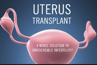
ASRM Video article: Technique of uterine procurement from a diseased donor for uterine transplant


Uterine transplantation is the final frontier in the quest to provide a child-bearing option for patients with absolute uterine factor infertility. This procedure can become a promising option for the approximately 1.5 million women worldwide for whom pregnancy is not possible because of the absence of the uterus or presence of a nonfunctional uterus.  Only recently we heard the news from Brazil about the first live birth following the uterine transplant from a deceased donor.

This informative and detailed video from the American Society of Reproductive Medicine (ASRM) outlines the key steps in the successful procurement of uterus from a deceased donor using illustrations and footage from a trial transplant.

The video also describes the advantage of deceased donor transplant over live donor transplant in terms of surgical time, ethical issues, larger availability and eliminating the risk of surgical complications in the live donor.


Monday, December 17, 2018

RCOG updates its guidance on Pregnancy pain relief


It is important to prescribe the correct analgesic during pregnancy to optimize the maternal and neonatal outcome. Inadequate pain-relief in pregnancy leads to anxiety and psychological strain for mother and affects her ability to provide care for her newborn–note the recent Scientific Impact Paper published December 12, 2018, in the BJOG.

The paper was developed for the Royal College of Obstetricians and Gynaecologists (RCOG) by a group of eminent doctors and researchers following the advisory issued by the Medicines and Healthcare products Regulatory Authority (MHRA) and European Medicines Agency (EMA) on the use of codeine in pregnancy because of neonatal mishap and three other fatalities in children.

The findings reflect current NHS guidance on the use of medical pain relief options in pregnancy and during breastfeeding. However, the guidance does not address the concerns for Intrapartum analgesia.
Women often need analgesic during the antenatal period for a headache, backache, and pelvic pain. 

Non-pharmacological interventions like relaxation, adequate rest, heat, massage, acupuncture, posture training, physiotherapy, and exercise should be considered before starting any drugs. Notably, all drugs should be avoided during early pregnancy to prevent the teratogenic effects of drugs during the organogenesis period (4-10 weeks).

Paracetamol
Paracetamol remains the drug of choice for pain relief throughout pregnancy, although few studies have demonstrated an increased incidence of childhood asthma, behavioral problems, and a delay in gross motor and communication development in children with long‐term antenatal exposure.

NSAIDs 
Studies have failed to demonstrate any clear association between congenital malformations and the use of nonsteroidal anti-inflammatory drugs (NSAIDS) in early pregnancy.

However, there is conflicting evidence regarding the use of NSAIDs and risk of miscarriage in the first trimester.  Therefore, NSAIDs be avoided during pregnancy, especially during the first trimester. They should only be used when absolutely necessary in conditions such as a migraine or ankylosing spondylitis, that too in lowest possible dose and for the shortest duration of time.

Use of NSAIDs should be avoided after 30 weeks of gestation because it can cause neonatal pulmonary hypertension and premature closure of the ductus arteriosus. NSAIDs also reduce fetal renal blood flow leading to decreased urine production and causing oligohydramnios. 

Opioids (including codeine, dihydrocodeine, tramadol, and morphine)

The review says that opioid analgesic should only be used under the care of health professional in the lowest possible dose and shortest amount of time. It should be used when paracetamol has been ineffective. Limited data available does not point towards increased risk of fetal toxicity.

However, if used around the time of labor, it may lead to neonatal respiratory depression and if used for prolonged time cause neonatal withdrawal symptoms and maternal dependence.

The guidelines also highlighted the critical difference between the use of codeine and DHC during breastfeeding. If stronger analgesia is required, Dihydrocodeine (DHC) is much safer to use as compared to codeine which causes fetal toxicity.

Low doses of dihydrocodeine in combination with paracetamol can be bought over the counter but regular use of any opioid beyond 3 days should be under close medical supervision.

Gabapentin
Gabapentin is mostly used in chronic pain syndrome, and only a few studies have been done on the use of gabapentin in pregnancy. Evidence does not link the use of the drug to a particular birth defect; however, women are advised to take high doses of folic acid before conception and during the first trimester.

Gabapentin use in pregnancy after 30 weeks is very limited, and if used around the time of delivery it may lead to neonatal withdrawal syndrome.

Analgesics at the time of discharge
Majority of women who require analgesics at discharge should be prescribed paracetamol or ibuprofen. If stronger analgesics are required (cesarean delivery), women should be sent home with a limited supply of DHC. Tramadol is only given if a woman is intolerant to DHC.

Dr Dina Bisson, consultant obstetrician and gynecologist at the North Bristol NHS Trust and the lead authors of the guidance said: "It is absolutely essential that pain is managed appropriately during pregnancy and breastfeeding. Many women may develop headaches, lower back pain, and pelvic pain during pregnancy and breastfeeding, while others may have chronic conditions, where pain management is necessary.

"If pain is not adequately managed, this can have a negative impact on a woman's physical and mental wellbeing.


Friday, December 14, 2018

Higher aerobic fitness linked to longevity and better quality of life


A new study from Cleveland clinic found that increased cardiorespiratory fitness (CRF) or aerobic fitness is associated with lower long-term all-cause mortality with no upper limit observed for the amount of exercise. Patients who demonstrated extreme CRF (≥2 SDs above the mean for age and sex) was at the lowest risk of adjusted all-cause mortality as compared to other performance groups.

People with poor aerobic fitness face the same likelihood of dying as that due to chronic diseases like diabetes, hypertension and cardiovascular disease note the researchers of this retrospective study published in the Journal of the American Medical Association Network Open.

This retrospective study recruited the largest reported cohort of 122 007 patients between Jan. 1, 1991, and Dec. 31, 2014, who underwent Exercise treadmill testing (ETT) at a tertiary care center. ETT eliminates the bias introduced due to self-reporting and is the most widely used method to measure the CRF.

Demographics and other co-morbidities were noted at the time of undergoing specified stress test under the supervision of exercise physiologist. Patients were classified into performance group by age- and sex-associated cutoffs as follows: elite (≥97.7th percentile), high (75th-97.6th percentile), above average (50th-74th percentile), below average (25th-49th percentile), and low (<25th percentile).

The mean age was 53.4years, and about 60% were male. As the performance level increased, the associated co-morbidities decreased except for hyperlipidemia (present in 30% of elite performers).

After multivariate analysis, it was seen that at any given point of time, patients in the elite group were 80% less likely to die as compared to patients with low CRF (elite vs. low: adjusted HR, 0.20; 95% CI, 0.16-0.24; P < .001). The survival benefit was more evident in older patients (70 and more) and those with hypertension. Long-term survival was adjusted for a patient’s age, sex, height, weight, BMI, medications, and comorbidities.

Hypertensive patients in the elite group were 30% less likely to suffer mortality as compared to those in the high-performance group (75th-97.6th percentile).

Patients with lowest aerobic fitness (<25th percentile) faced risks that were comparable or more than patients with chronic disease like CVD (24% more), diabetes (40% more) and smoking (40% more). 

These findings highlight the importance of aerobic fitness concerning prolonging life and improving the quality of life. This study reinforces the results of previous studies that have demonstrated the benefits of achieving CRF.

“Aerobic fitness is something that most patients can control. And we found in our study there is no limit to how much exercise is too much,” said Wael Jaber, M.D., Cleveland Clinic cardiologist and senior author of the study in a news release.  “Everyone should be encouraged to achieve and maintain high fitness levels.”

It also stresses the importance of continued high-level physical activity in older adults and the benefits of maintaining the highest aerobic fitness. Some recent studies have demonstrated an association between extreme high level of exercise and an increased incidence of atrial fibrillation, coronary artery calcification, myocardial fibrosis, and aortic dilation. However, it is not clear whether these findings are a simple physiological adaptation or because of cardiovascular pathology.

“We were particularly interested in the relationship between extremely high fitness and mortality,” said Kyle Mandsager, M.D., an electrophysiology fellow at Cleveland Clinic and the lead author of the study.  “This relationship has never been looked at using objectively measured fitness, and on such a large scale.”

The authors conclude, “Cardiorespiratory fitness is a modifiable indicator of long-term mortality, and health care professionals should encourage patients to achieve and maintain high levels of fitness.”

Wednesday, December 12, 2018

Menopausal transition period is a "critical window" for cardiovascular prevention in women


A healthy lifestyle during the perimenopausal and menopausal years reduces the risk of cardiovascular events in later life report the result of a prospective, cohort study published in in the December 4 issue the Journal of the American Heart Association.

The study is a secondary analysis of data from the ongoing, multicentric, multiethnic, prospective Study of Women's Health Across the Nation (SWAN) initiated in 1996 to know more about transition across menopause.  The researchers looked at data from 1143 women to create a composite 10-year average Healthy Lifestyle Score (HLS) involving smoking, diet quality, and physical activity. All three are modifiable behavioral risk factors for CVD and earlier studies have shown an inverse association between healthy lifestyle and various CV outcomes.

The study participants were followed for an average of 15 years with the last follow-up in the year 2015-2016. Carotid ultrasound scans were performed after 14 years to measure the markers of subclinical atherosclerosis which include common carotid artery intima‐media thickness (CCA‐IMT), adventitial diameter (CCA‐AD), and carotid plaque.

Information about the diet was collected at baseline, visit 5, and visit 9 using a modified version of the Block Food Frequency Questionnaire (FFQ) and the amount was quantified using the Alternate Healthy Eating Index (AHEI).

To assess whether the recommended physical activity (≥150 minutes/week of moderate-intensity physical activity) is met or not, the participants were asked to fill the sports and exercise questions on the Kaiser Physical Activity Survey.

Data on smoking were collected using the standardized questions from the American Thoracic Association.

The HLS was calculated based on the sum of individual scores on all the three parameters at baseline, visit 5 and visit 9 and averaged to get the final score. Other covariates included in the study at baseline were age, race/ethnicity, education, financial status, marital status, depression, self-reported health status, and menopausal status.

Physiological risk factors, including BMI, high blood pressure, impaired fasting glucose, serum triglycerides, total cholesterol, HDL cholesterol, LDL cholesterol, use of antilipidemic medications, and use of antihypertensive medications were adjusted in a separate model.

The association between the individual component of HLS and the three markers of subclinical atherosclerosis were also looked at.

At 10 years follow-up, average HLS was found to be inversely and statistically significantly associated with CCA-IMT and CCA-AD — an association that persisted, even after "extensive" adjustment for confounders (P = .0031) and physiological risk factors (P < .001 for both CCA-IMT and CCA-AD).

Compared with participants with the lowest range of HLS, those with highest HLS had a 0.024 mm smaller CCA-IMT and a 0.16 mm smaller CCA-AD.

After adjusting for various physiological risk factors, the researchers did not find a statistically significant association between carotid plaque and average HLS.

Analyzing individual component of HLS, smoking conferred the highest risk for a CV event and those who never smoked had a 0.047 mm smaller CCA-IMT, a 0.24 mm smaller CCA-AD, and 49% lower odds of having a higher carotid plaque index.

The investigators message for the physicians “the menopausal transition represents a crucial, yet understudied, window of increased cardiovascular risk in women. For the prevention of future cardiovascular disease among women undergoing the menopausal transition, the physician should focus on modifiable health behaviors including smoking, diet, and physical activity."


Tuesday, December 11, 2018

ASRM Video article: Hysteroscopic removal of IUD in pregnancy


The use of Intrauterine Devices (IUD) has increased in recent years. Used correctly, the failure rate for IUD is less than 1%. If a patient gets pregnant with IUD in place, the risk of adverse outcomes like spontaneous abortion and preterm birth increases many-fold.

WHO advises removal of IUD at the earliest gestational age possible if strings are visible on vaginal examination. If strings are not visible there are 3 ways to manage a case of IUD with pregnancy:
Expectant management
Removal with a grasper under ultrasound guidance
Removal under hysteroscopic visualization


Here is a recent video-article published in December issue of Journal Fertility and Sterility, showing step by step procedure of hysteroscopic removal of IUD in 4 patients. In all four cases, the strings were not visible during a vaginal examination. After removal of IUD, all patients had uneventful live term delivery.


All patients were informed about the risk of the procedure, although negligible–– including rupture of gestation sac and pregnancy loss, consent obtained, viability and location of IUD confirmed by pre-op ultrasound and prophylactic antibiotics given. The actual procedure includes:
Vaginoscopic hysteroscopy
IUD localization with or without imaging guidance
Removal of the IUD with the help of hysteroscopic grasper

The key is to use slender hysteroscope and use the minimal amount of distention media. The method is safe with minimum procedure-related complications and > 90% ongoing pregnancy rate.

Here is the American Society for Reproductive Medicine (ASRM) video showing step by step procedure of hysteroscopic removal of IUD in 4 patients. 


Monday, December 10, 2018

Hologic launches multipurpose Omni-Hysteroscope


Hologic announced the launch of its FDA cleared, innovative 3-in-1 Omni™ Hysteroscope equipped with advanced visualization capabilities to streamline the diagnostic and therapeutic procedures.

The scope comes with 3 interchangeable sheaths which eliminate the physician need to change the scope between diagnostic and treatment procedures. Additionally, the scope is armed with Premium MyoSure optics to ensure clarity and quality visualization throughout the procedure.

The patients experience less pain and discomfort because the scope can be easily inserted because of thin sheaths (3.7 mm diagnostic sheath; 5 mm operative sheath; 6 mm operative sheath). It also provides long 200 mm working length to efficiently work with obese patients.

The sheaths are compatible to use with Hologic’s Fluent™ hysteroscopic fluid management system and all MyoSure® tissue removal offerings including MyoSure REACH, MyoSure XL, MyoSure LITE, and MyoSure MANUAL devices.

“Experts agree that direct visualization of the uterine cavity in women with abnormal uterine bleeding is the gold standard that allows physicians to accurately identify and collect quality samples and remove pathology – in a safer and more effective manner than blind biopsy and curettage,” said Edward Evantash, M.D., Medical Director and Vice President of Global Medical Affairs, Hologic. 

“Featuring three easily interchangeable sheaths in one scope, our new Omni scope gives physicians excellent visualization capabilities with the convenience of seeing and treating pathology in a more streamlined procedure,” he added.  

Saturday, December 8, 2018

First live birth following a uterine transplant from a deceased donor


A Brazilian woman becomes the first person to successfully give birth to a healthy baby after receiving a uterine transplant from a deceased donor. This comes after a history of previous 10 unsuccessful attempts at live birth after deceased donor transplants in the United States, the Czech Republic, and Turkey.

This is a milestone in the history of uterine transplants as it opens the path forward for achieving successful pregnancies without the need of live donor and donor surgery. The details of the case were published by the Lancet on December 4, 2018, accompanied by an editorial.

Dr. Dani Ejzenberg, PhD, the lead author and researcher from the Department of Obstetrics and Gynecology, Hospital das Clínicas, Faculty of Medicine, University of São Paulo, said in a press release by Lancet, "The use of deceased donors could greatly broaden access to this treatment, and our results provide proof-of-concept for a new option for women with uterine infertility."

Ejzenberg and colleagues report that the 32-year old woman was born without a uterus because of congenital uterine agenesis, also called as Mayer-Rokitansky-Küster-Hauser (MRKH) syndrome, which affects 1 in 4500 women.

The woman received uterus in September 2016 after 10.5-hour surgery from a 45-year old multiparous woman who died of subarachnoid hemorrhage. After the transplant, the woman was discharged on the 8th postoperative day with immunosuppression achieved and maintained with prednisolone and thymoglobulin and continued via tacrolimus and mycophenalate mofetil (MMF) when at five months post-transplant, azathioprine replaced MMF.

The woman got her first period 37 days after transplant and after that continued to have regular cycles every 26–32 days.  Pregnancy occurred following single embryo-transfer after seven months from cryopreserved blastocyst from a successful in-vitro fertilization four months before the transplant surgery.

The pregnancy progressed uneventfully and was monitored by ultrasound and Doppler flow velocity waveforms of uterine arteries, fetal umbilical, or middle cerebral arteries.

A healthy, female baby weighing 2550 g was delivered by elective cesarean section at 36 weeks on December 15, 2017. At the time of writing the article, the baby is healthy and developing normally.

The uterus was also removed at the time of cesarean section, and immunosuppressive therapy stopped.

Cesar Diaz-Garcia, MD, Nuffield Department of Women's and Reproductive Health, University of Oxford, UK, and Antonio Pellicer, MD, PhD, Department of Pediatrics, Obstetrics and Gynecology, University of Valencia, Spain, writes in an accompanying editorial, "In a context in which the scarcity of human data is still the norm, [the report] reveals a breakthrough in the field of uterus transplantation."

The first uterine transplant from a living donor took place in Sweden in 2013, followed by first live-birth after transplant in 2015. Since then, 11 more births have taken place worldwide, albeit all from live donor transplant.

However, uteri from living donors are limited because the person has to a relative of the recipient, thereby, limiting the option for women with uterine factor infertility. If uterine transplant after dead donor becomes successful, uteri will not be in short supply as people are more willing to donate organs after death than when they are alive.

The authors agree that many potential issues need to be addressed before deceased donor transplant becomes a norm. These include standardization of operative procedures, immunosuppressive therapy before and during pregnancy, criteria for rejection if it happens, and long-term outcome of the baby delivered after transplant.